I haven’t posted in a while because I went on vacation and lost my momentum for a bit after some “long haul Covid” symptoms started flaring up again. Fasting and high dose vitamin D seems to help me the most. I’ve only tried sodium citrate once at a low dose but intend to develop a proper protocol for it soon. I’m having a lot more brain fog than usual lately, so this post will be mostly pictures with less analysis.
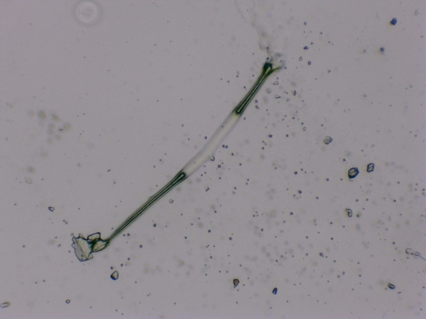
The following photos are from a saliva sample that was initially taken on 5/25, so about 3 weeks have passed since then. Many other researchers have noted that it can take weeks or even months for telltale signs of the nanotech to start showing up on a slide. In my last post, I noted the development of filament networks that one commenter noted are a bit plant-like in appearance. The filaments and tubes seem to be the product of edges of hydrogel sheets merging together.
Here is a quick video of the hydrogel sheets forming what may be a type of tube or filament. They look pretty similar to what I was seeing in my first post with the blood sample and tubes that reminded me of antennas.
If you want to better understand the dynamics of the hydrogel sheets forming into these tube-like structures, there’s a great discussion on Rumble between Drs. Ana Mihalcea and Sandy Corlett. Dr. Corlett has also seen these filament-like structures in the blood and believes it happens when the edges of two hydrogels meet and their combined material forms a hollow tube. You can watch the full interview on Rumble if you follow this link and their discussion of these antenna-like tubes starts at about the 1: 03 (one hour, three minute) mark.
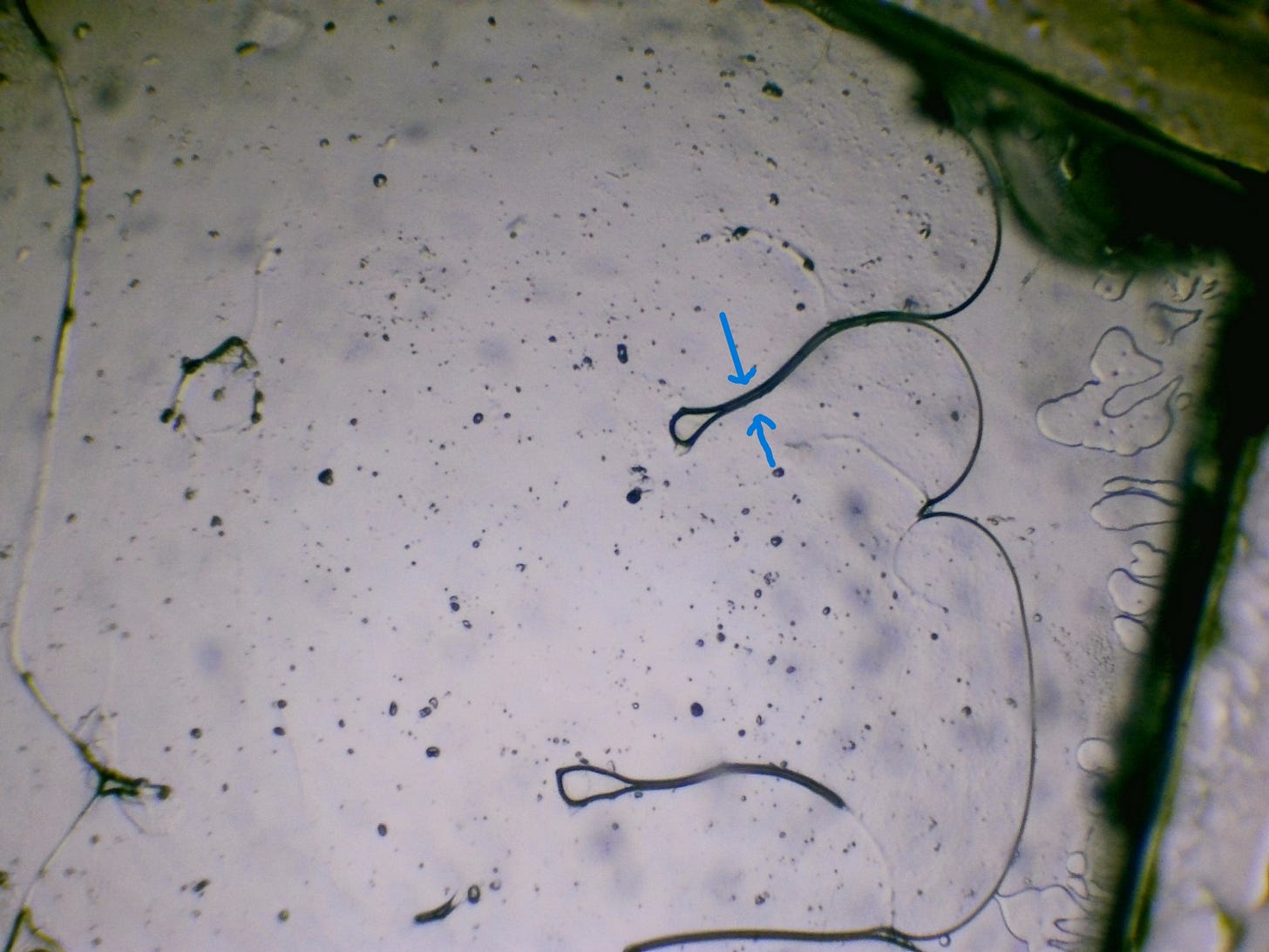
Take note of where the two blue arrows meet. It seems that two different surfaces of the hydrogel are merging together and the edges of the hydrogel itself are forming the tubes, filaments, ribbons and other structures. If I remember correctly, Ana Mihalcea recently showed strong evidence that the ribbons are formed from the gel and not the other way around. I’ll go through that research again when I have the time.
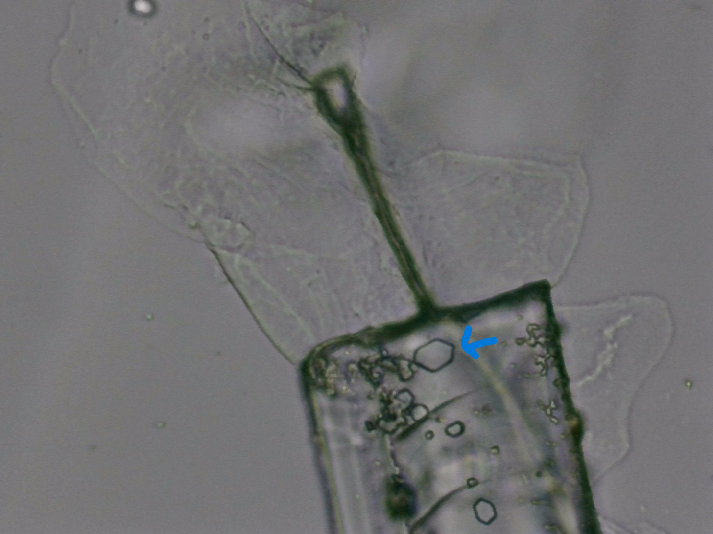
I also noticed the beginning stages of chip-like structures, which typically present as a rectangular shaped crystal attached to a ribbon. I didn’t see these at week one and week two. I drew a blue arrow to point out some epithelial cells from the lining of my cheek. It appears that the hydrogel may be trying to siphon materials from whatever it encounters in its immediate environment to help build the chips, ribbons and other structures.
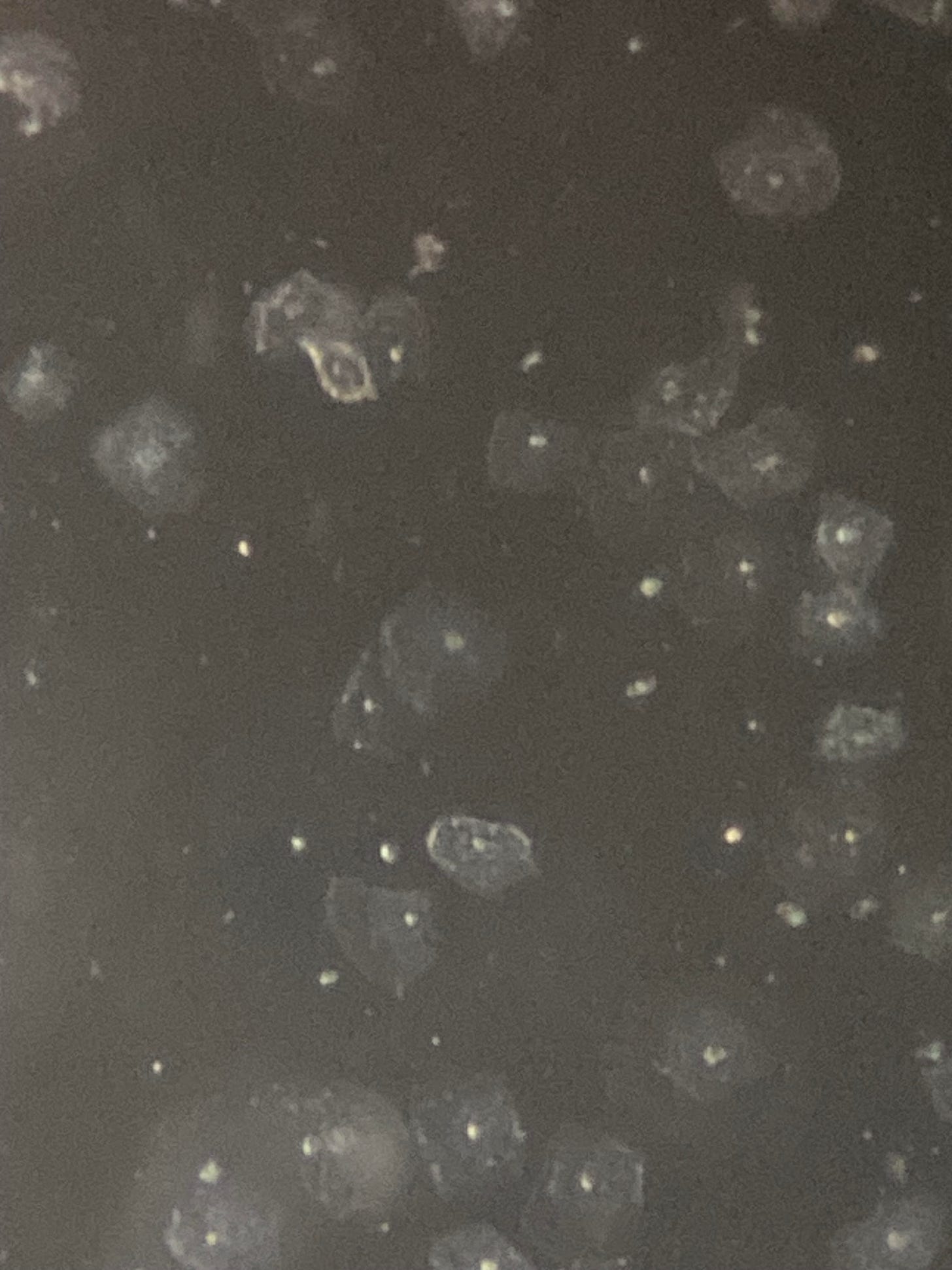
For reference, here’s a photo of just the epithelial cells by themselves. This image is from the first day I took the sample, probably under 60 seconds from the moment of putting the drop of saliva onto the slide. Epithelial cells form the inner lining of most of your organs and cavities, including the lining of your mouth. Basically just like the skin or epidermis, but on the inside instead of the outside of your body.
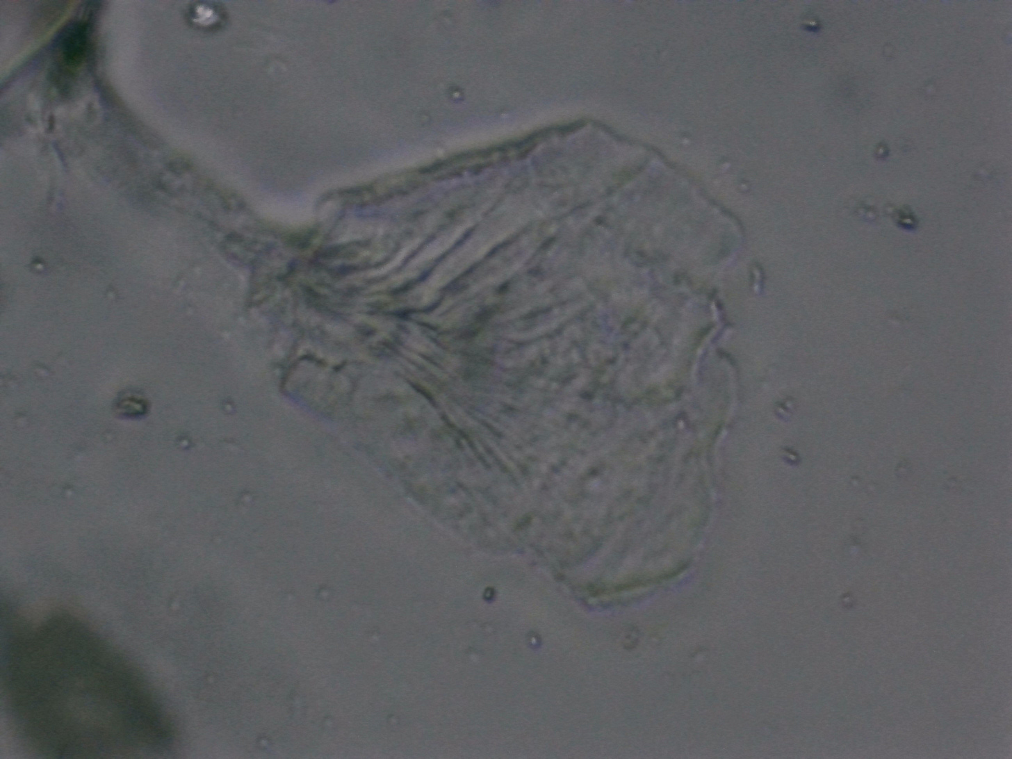
And here’s a closeup of an epithelial cell being grabbed by a nearby filament or ribbon. It almost looks like the tube/filament is sucking the materials out of the epithelial cell like a straw.
Apologies for the dark and blurry images, but I’m doing the best I can. Here’s a video showing a bit more depth and contrast.
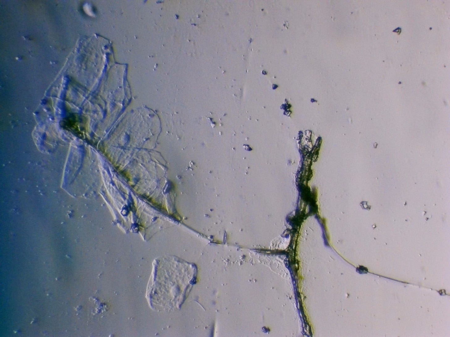
Here’s a larger clump of epithelial cells being pulled towards or sucked into the filament tubes. It’s hard to tell if the tubes are creating a suction or if the cells are just getting dragged along in the process of the hydrogel sheets merging to form the tubes.
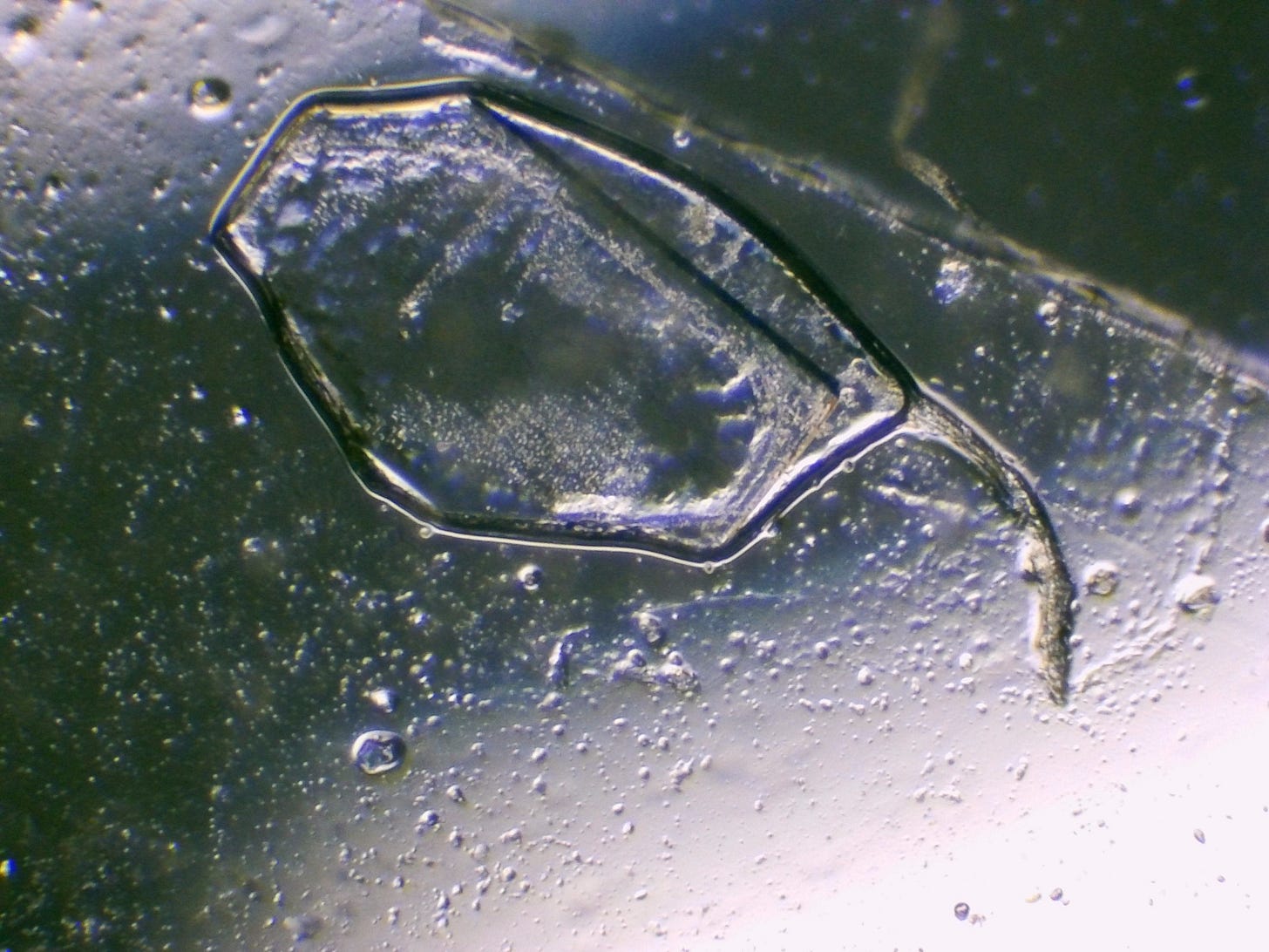
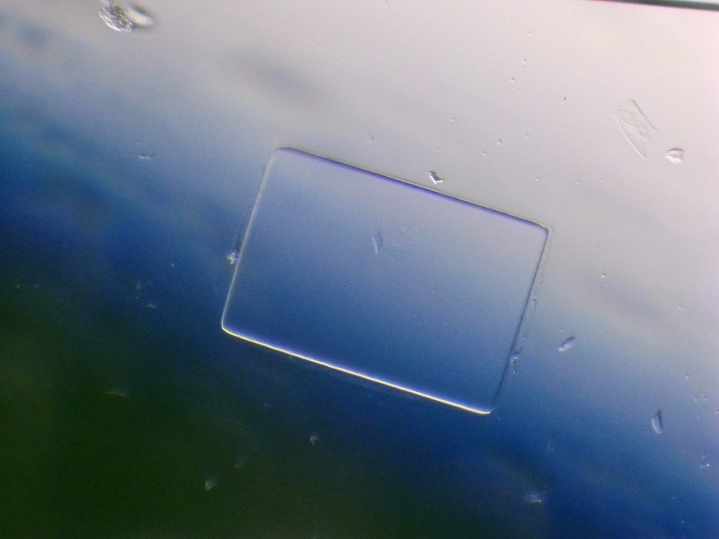
Here’s a closeup of the “chip” or chip-like structures. They almost look like little razorblades :(
It’s not particularly fun to think about what these structures might be doing to our bodies and our health.
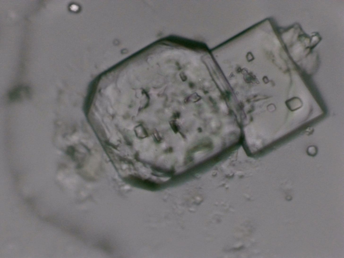
I think it’s worth pointing out some hexagonal shapes that seem to be inside the chip (indicated by blue arrow). I was chatting with a group of nano researchers and someone mentioned that they had seen flat hexagons that normally appear by themselves and not in the immediate vicinity of chips or ribbons. I also saw standalone flat hexagons recently develop in an albuterol sample, which I will discuss in the next post.
Here’s a broader look at the whole filament network that I first showed in the last post. It has evolved a little bit from the last post, but not a whole lot. Someone mentioned that it looks a bit like plants. It sort of looks like flowers and vines to me as well. The epithelial cells scrunch up while getting pulled towards the tubes and look a little bit like petals.
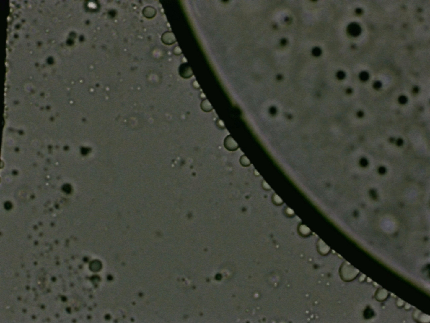
Here’s a hydrogel formation with what Ana Mihalcea calls micellar cells inside of it. David Nixon and Karl C. call them constructor bubbles. I’m still trying to decide which terminology to use.
I recently discovered the work of Clifford Carnicom and the Carnicom Institute and heard him mention in an interview something about the formation of bubbles along the inside and/or outside surfaces of larger (hydrogel) bubbles. I haven’t yet done a deep dive into his work, but I did take note of ridges of bubbles in a line. I believe Karl C. did a dive on this topic as well, and I may try to do a deeper dive on it at some point.
Closer look at a straight edge of hydrogel/graphene. You can see it folds over into a flap in the second photo.
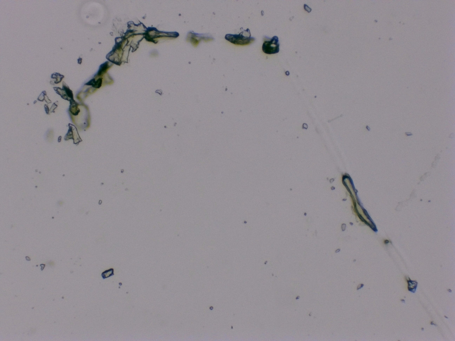
Here’s a closeup look of some tubes that seem to break off and restart in different places. It almost looks like faint light is emitting from where the tube should continue to look solid but becomes transparent.
I’m not sure what this next image is, but it’s interesting.
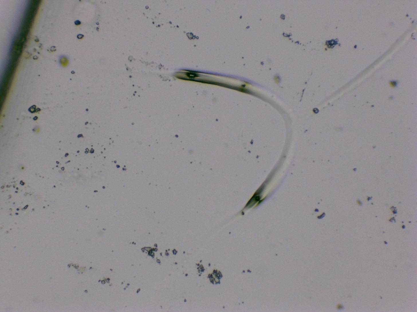
It’s interesting how some of the tubes seem to inexplicably disintegrate and fragment while other tubes grow stronger and more complex. This next tube is almost fully transparent.
And here’s a broader look at a network of seemingly transparent tubes.
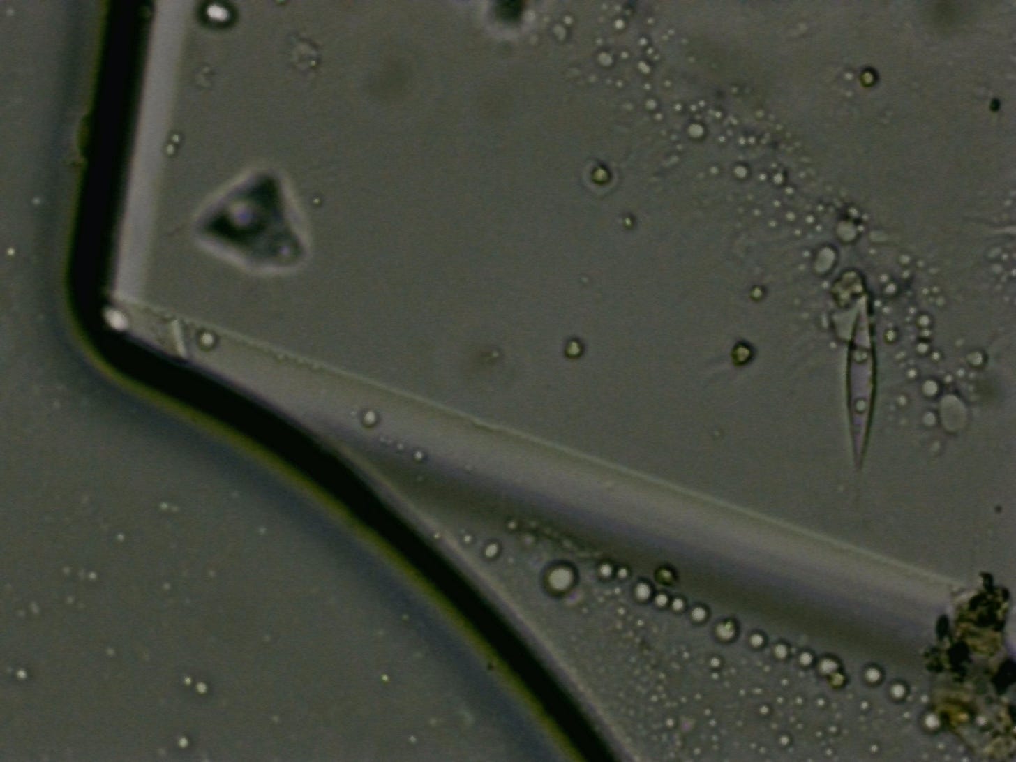
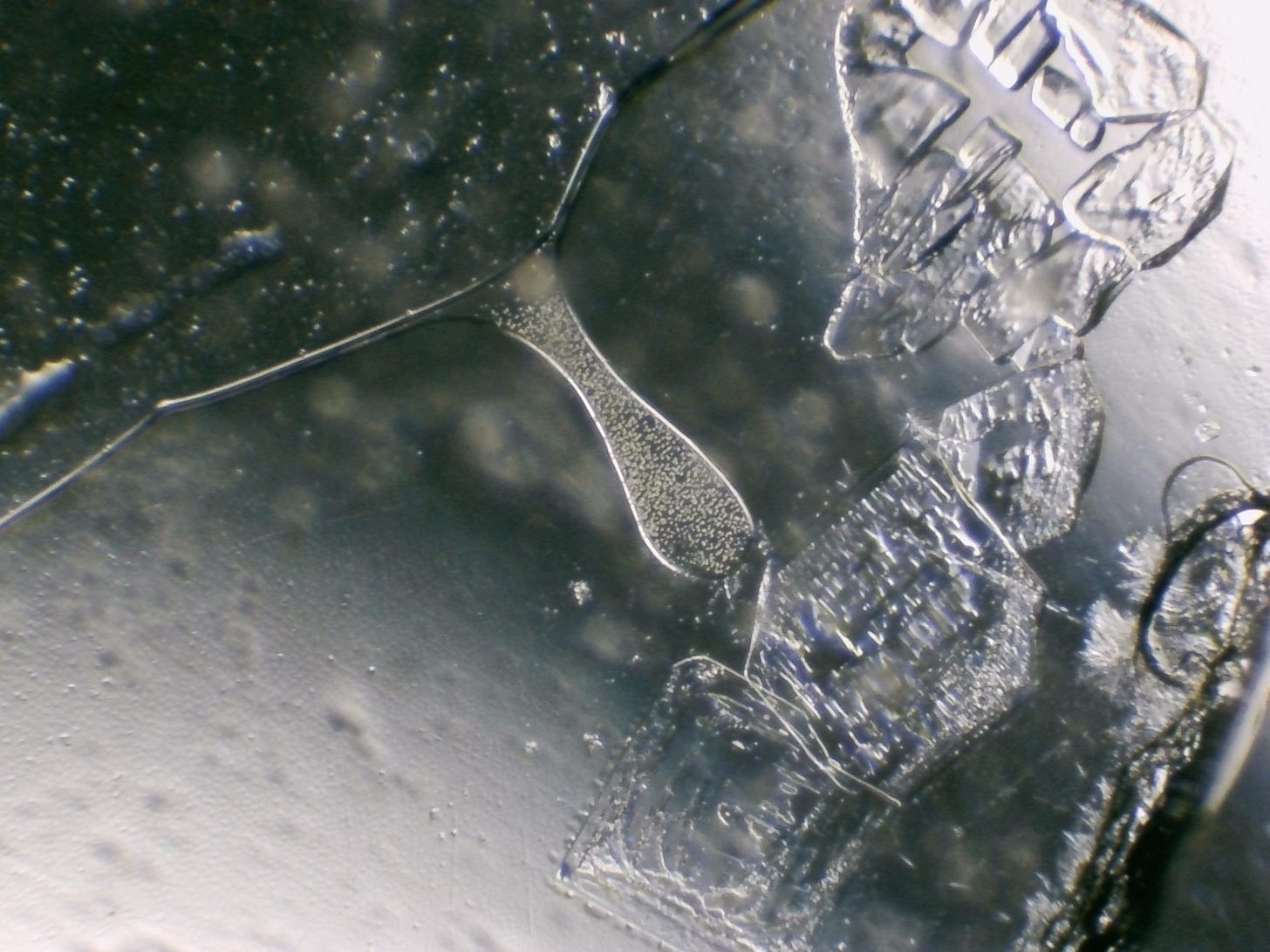
The next series of images were taken from the edge of the slide. I’ve noticed that there’s usually more action at the edges of the slides. There’s especially more crystal formation at the edges. You can see that here along with a teardrop-shaped hydrogel plate full of smaller micellar bubbles.
I thought this particular ribbon was interesting because it has horizontal bands of color and tapers at the end like a shoelace.
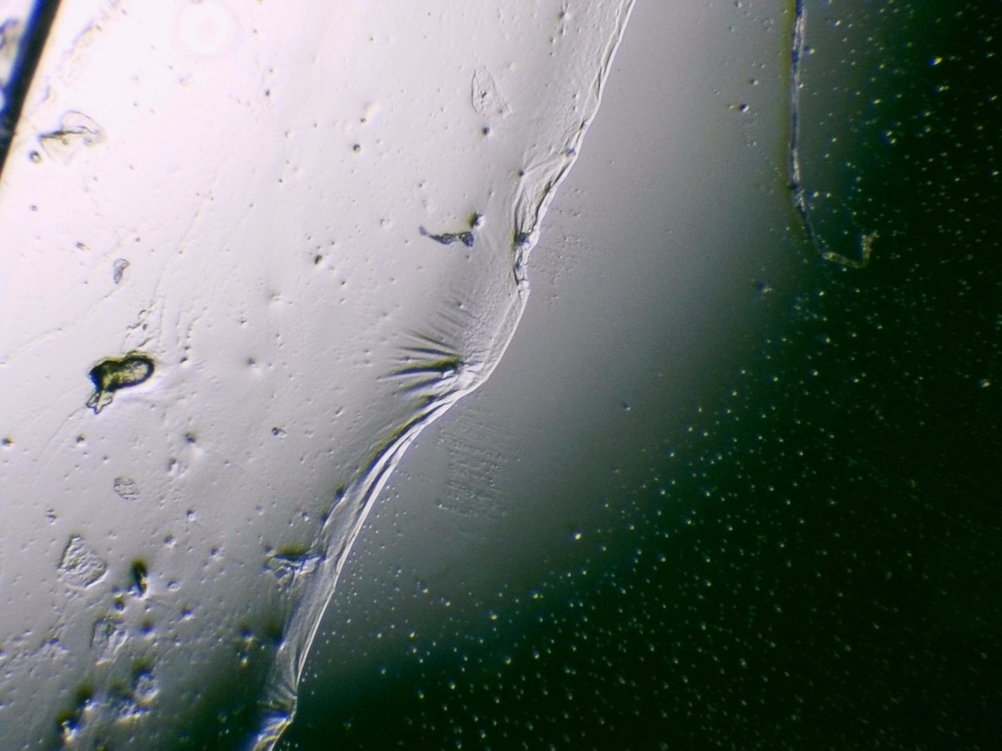
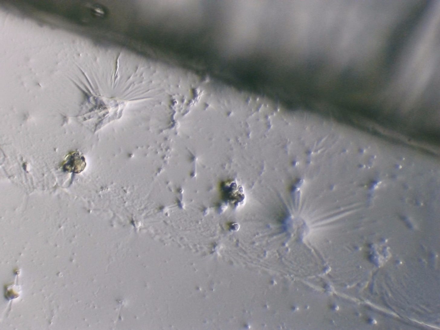
I noticed a sheet of hydrogel that seemed to be drying out or getting stretched in a way that reminds me of a sheet pulled taut and tight, creating a ribbed texture. I tried to get photos under different lighting so you can see the texture of the sheet.
It almost looks like the shoreline of a beach.
The last thing I observed was hard to explain and was forming at the edge of where the coverslip meets the slide. It seemed to have a metallic sheen with symmetrical formations of dots. It deserves it’s own deep dive so I will save that for a later post and just leave you with this one last video. Apparently this post is now too long to be emailed out to subscribers and so next time I’ll try to divide it up into smaller posts.
Hope you enjoyed the content! I’ll add a paid subscriber option soon if you’d like to donate. Thanks for the support.











You might want to try taking the master antioxidant- glutathione. It works wonders.
So sorry for the brain fog. It has been twenty years for me, so I know what torture you will be feeling - thanks for the images.