quick update
short post, minor updates
I’m so glad I took up this new hobby of microscopy. I also enjoy looking at the new space images coming from the James Webb telescope and I’m surprised at how similar looking up at the macro feels to looking down at the micro. I now fully understand why some call us microscopists “micronauts”.
The evolution of the budesonide covered in previous posts has slowed down quite a bit, though it is still changing from day to day. I will wait until I see some notably large developments before I post more pics. I also didn’t notice much change in the first blood samples. Going forward, just assume that no significant developments were noted if I don’t follow up on previous posts.
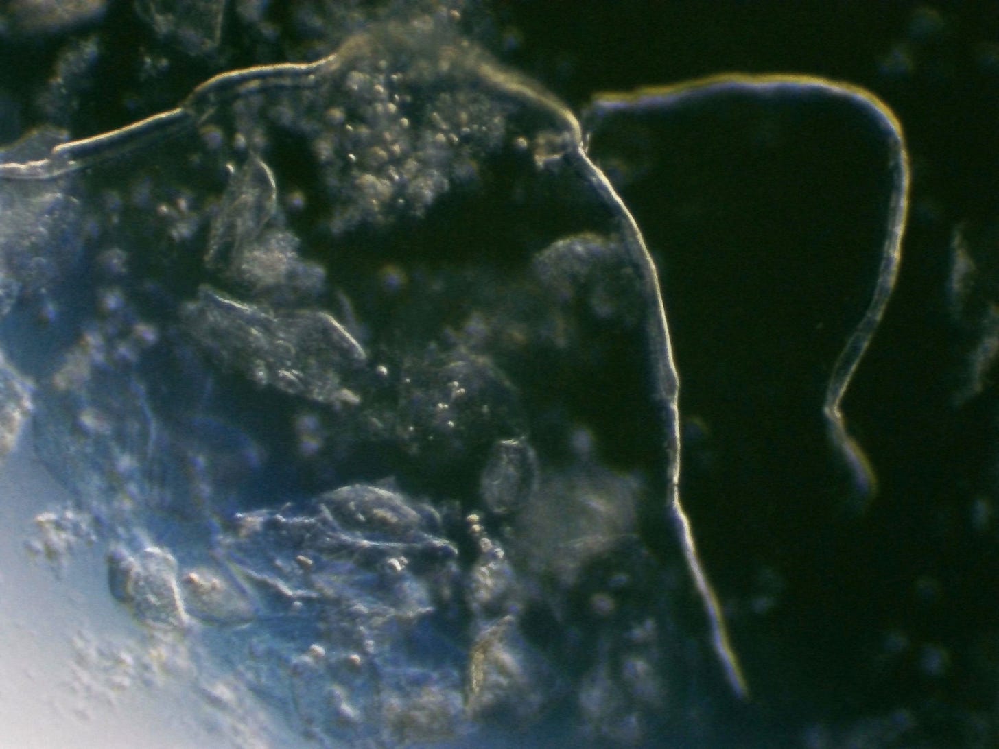
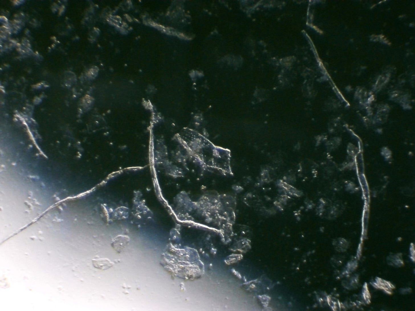
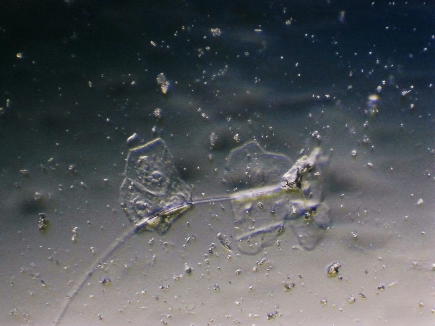
I did put a new sample of my saliva onto a plastic petri dish yesterday. It looked normal. Looking again now, roughly 24 hours later, so many ribbons have appeared that were not there yesterday. The vast majority of the material is squamous epithelial cells from the lining of my mouth. Those would be the transparent irregularly shaped cells with a nucleus in the center.
Please bear with me as I continue to refine my “DIY darkfield” technique. It looks like I may be able to install a separate adaptor for my Amscope that doesn’t come equipped with filter holders, but it will take some time for me to research and eventually purchase.
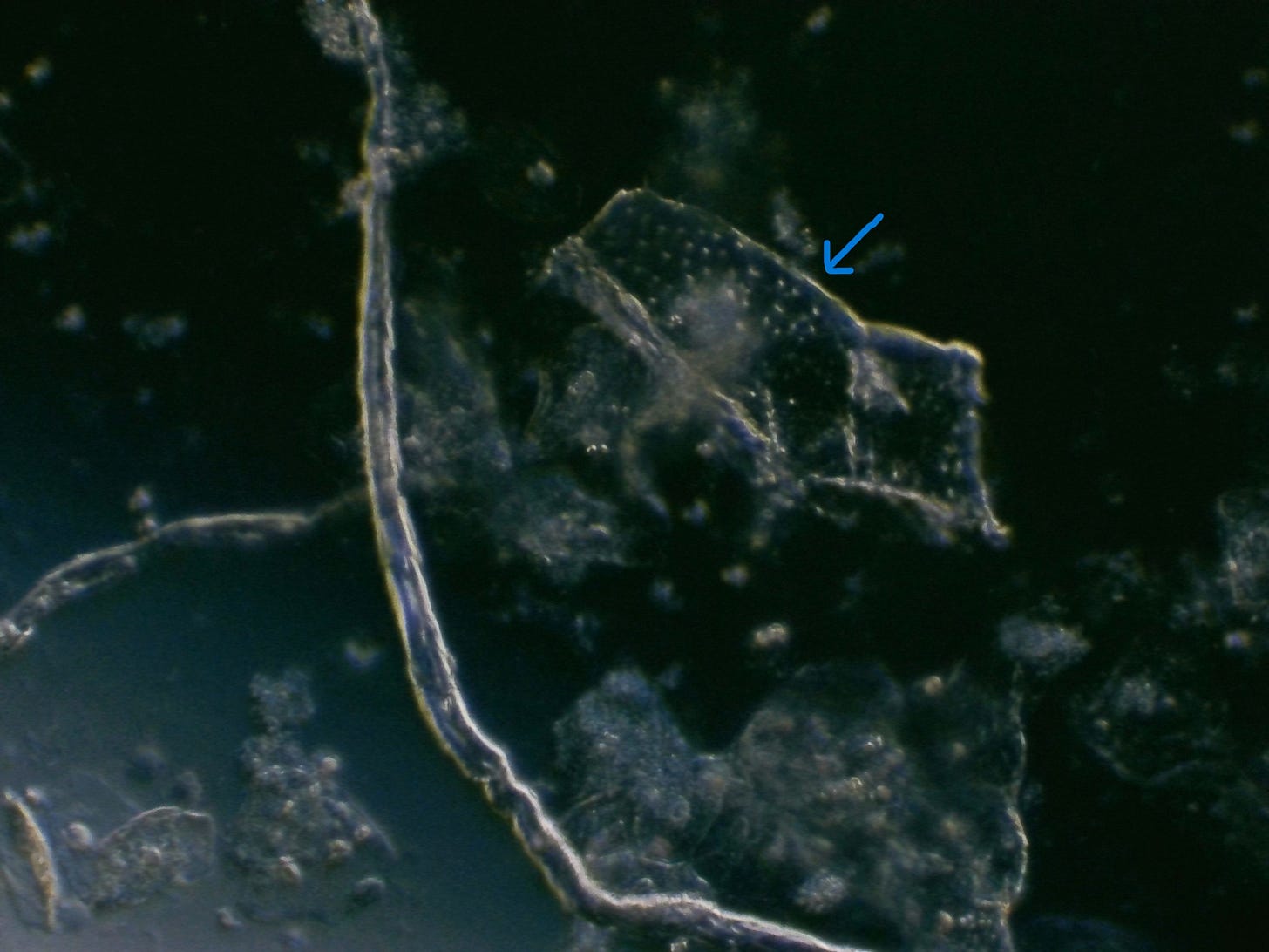
There was one object I noticed that may be a graphene sheet with “q dots” inside, indicated by the blue arrow. Because it was open petri dish, I didn’t feel comfortable using a higher objective that might get too close and accidentally touch the sample, so I didn’t really get a close enough look to see more detail. Please leave a comment if you have any ideas about what the transparent sheet with dots might be. It may be part of the lining of my mouth like the other squamous cells. Its proximity to the ribbons leads me to think it may be graphene.
That’s about all I have for now. The ribbons in the saliva that appeared literally overnight were innumerable and too many to count. I was surprised that so many could sprout up so quickly. I wonder if they were initially there the day before but just too small to see as they hadn’t grown yet. Perhaps isolating the saliva outside of my body at room temperature somehow has the effect of helping these ribbons grow. If you have any insight or theories, please drop it in the comments.
I’m slowing down a bit on microscopy over the next couple weeks as I’m about to take a vacation and much needed break from investigating the creepy crawlies. But I’ll try to get out at least one post a week. Thanks for reading and commenting.
Edit: haha, just kidding. Of course the situation had to escalate quickly to an insane level right before I was about to shut off my microscope and go to bed. I decided to put just one more slide on for a quick look before bed. I removed the petri dish with the large saliva sample and put on a slide of a drop of saliva under a cover slip, also collected yesterday. Again, with no notable irregularities visible on initial viewing about 24 hours ago.
But now there is a filament network already quite surprisingly complex that developed in a little over 24 hours. These filaments look a bit different to full “fibers”. They seem thinner, more gossamer, and are interconnected in branching patterns. There seem to be chip-like structures beginning to form, attached to or adjacent to the fibers in a typical presentation noted by many other researchers.
It is now 2:15am and I can hardly keep my eyes open, so I will end the post here. I have no explanation for why these branching filament structures would appear in a thin sample under a cover slip but not in the large sample in the petri dish. My only guess is that the thickness of the larger sample in the petri dish perhaps obscured these filaments if they in fact were present; or perhaps the thinness of the slide sample somehow facilitated the growth of these branching networks.
Off to bed. Will spend more time looking at these tomorrow.
Update 5/30: I just watched a discussion on Rumble between Drs. Ana Mihalcea and Sandy Corlett. Dr. Corlett has also seen these filament-like structures in the blood and believes it happens when the edges of two hydrogels meet and their combined material forms a hollow tube. You can watch the full interview on Rumble if you follow this link and their discussion of these antenna-like tubes starts at about the 1: 03 (one hour, three minute) mark.



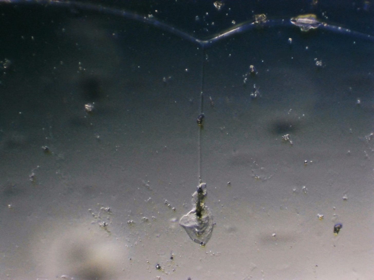
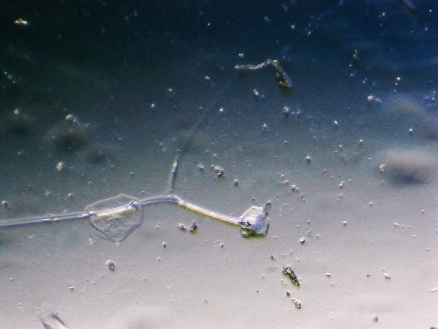
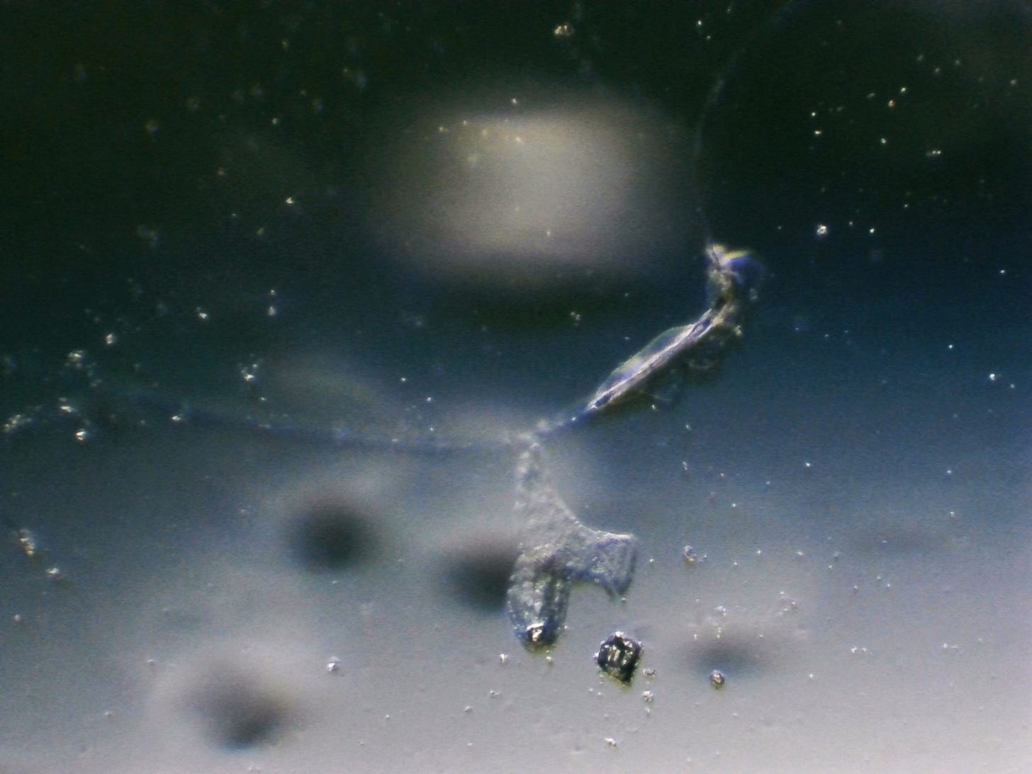
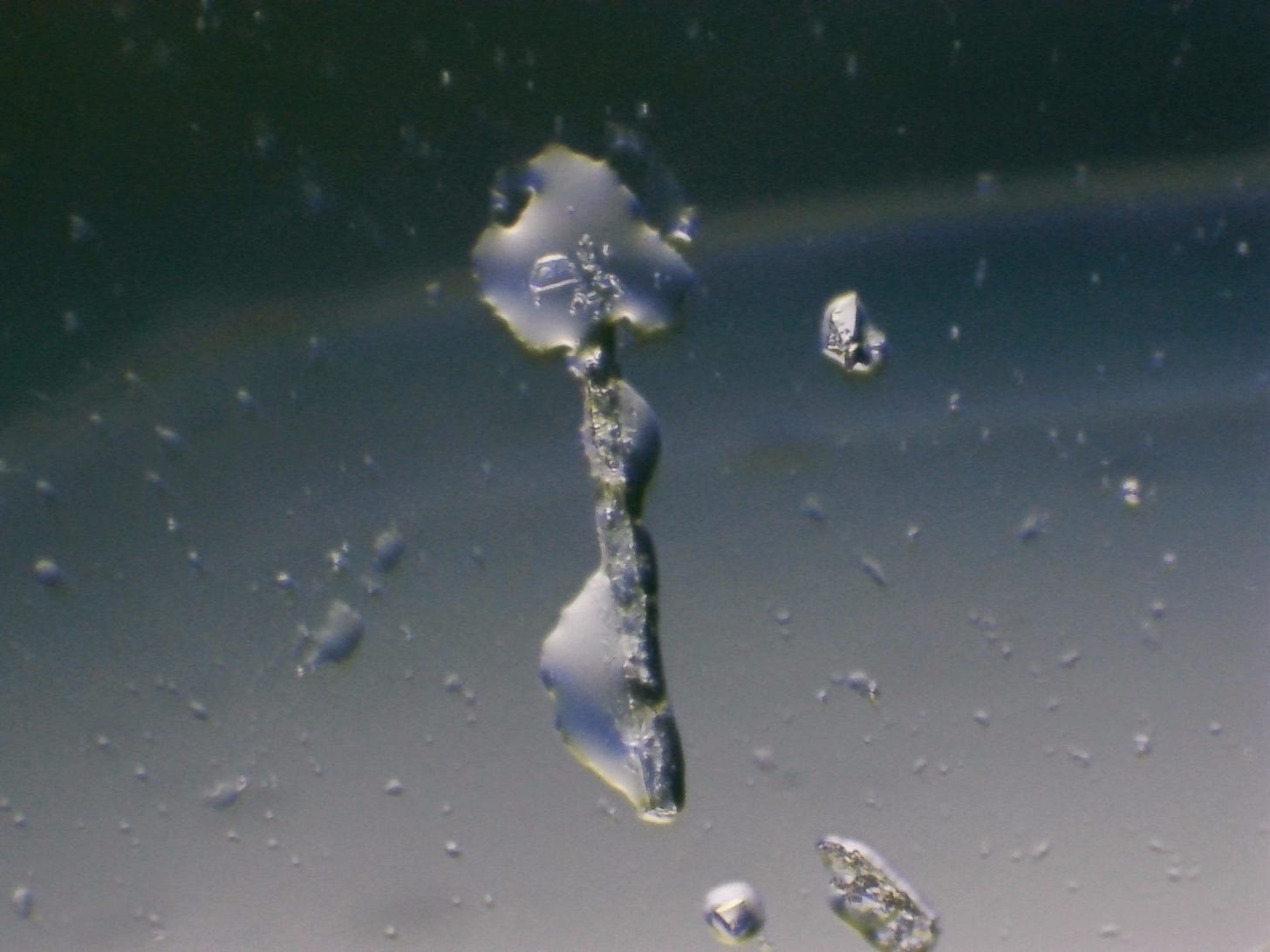

Hi there Michelle, wow, good captures. first of all looking at your fibres and how they presented…is a bit similar to FreedomWarriorWoman’s samples of a group of recent fibres resembling almost in a snake head at the termination on one end of her fibre samples (found in dental anaesthetic I believe). But yours are very different not seen any like this before, where the fibres seem to also come to a termination Head of a snake bulge - but then continues on from the snake head for another length of straight fibre to yet again another termination/end point and continuing on again in the thin thread length and also now branching off from the main fibre stem. An articulated fibre measured out in these bulges along its length creating segmented fibres with branches. Very odd.
Also it would have been good to have seen how those other puddle-like patches formed spreading out almost like leaves/puddles of gel? maybe from certain points along some of these fibres. You would have to have left filming video on a time-lapse to see how they had formed. Overall it’s a puzzling different expression of fibres you have caught for sure. Your still photos are really very clear and detailed and equally questionable features in there too. It’d be interesting if any other microscopists could recognise what those might be, again not looking familiar to me. Good catch, thanks for showing us.
Hi Michele~ The squarish thingy with textured dots that you think might be graphene, we in Dr. Nixon's group have seen in other things: Coffee and something else, can't remember. Not sure what it is – it might be a polymer mesogen or nanocrystal. La Quinta Columna will say that it is graphene, because they say Everything is Graphene. Interesting that you found in your saliva! Some of us called it a studded handbag. It belongs in the Nano Lexicon for sure!